
Figure 2.
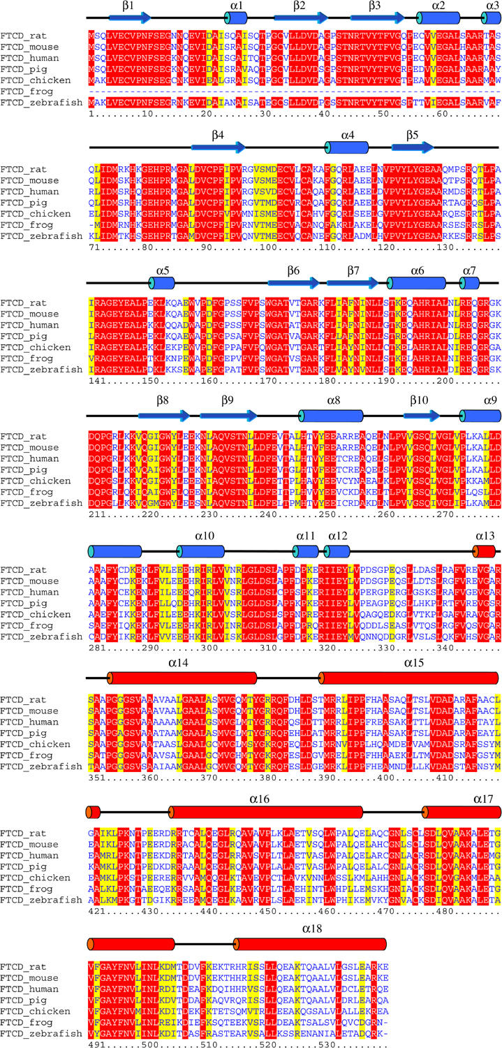
Secondary structure of rat FTCD monomer and multiple alignment of FTCD amino-acid sequences. The secondary structure elements of the FTCD crystal structure are drawn above the alignment with the rat protein sequence as reference, and those that are colored blue and red belong to the FT and CD domains, respectively. The alignment was obtained using the Clustal W program and colored by the Boxshade program. Only invariant and highly conserved residues are highlighted in red and yellow backgrounds, respectively.