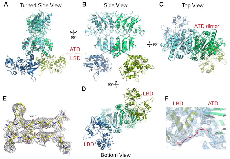
Figure 6. Structure of the symmetric dimers of rat GluD2 ectodomain.
A–D. GluD2 ectodomain dimers in four different views.
E. Representative 2mFo-DFc electron density from the beta-strands of the C-terminal lobe of the ATD domain at 1.0 σ.
F. NCS-averaged 2mFo-DFc electron density for the unmodeled ATD-LTD linker, which packs both on to the ATD and LBD.

