
Figure 3.
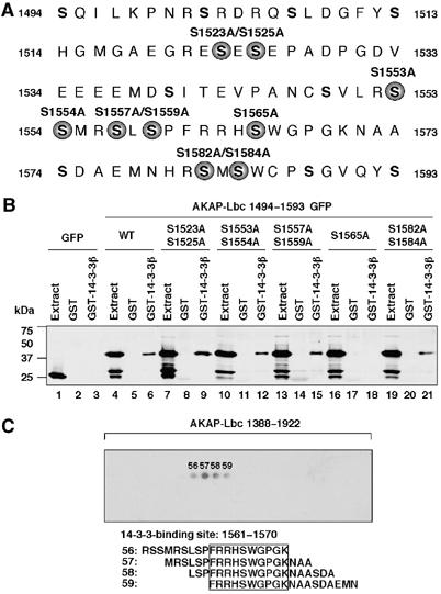
14-3-3 interacts with AKAP-Lbc through a phosphoserine-containing motif. (A) The region of AKAP-Lbc encompassing residues 1494–1593. Serine residues mutated to alanine are indicated by solid circles. (B) Extracts from HEK-293 cells expressing either GFP or GFP-tagged AKAP-Lbc fragments encompassing residues 1494–1593 carrying point mutations of various serines were incubated with glutathione-sepharose beads coupled to GST alone or to GST-tagged 14-3-3β. The GFP-tagged AKAP-Lbc fragments eluted from the beads were detected as indicated in Figure 2C. (C) Peptide array analysis of the 14-3-3-binding site on AKAP-Lbc. An array of overlapping 19-residue peptides spanning the region of AKAP-Lbc included between residues 1388 and 1922 was submitted to PKA phosphorylation and incubated with 50 nM S-tagged 14-3-3β. Solid phase binding was assessed with HRP-conjugated S-protein. The 14-3-3β-binding peptides are numbered. The results are representative of three independent experiments.