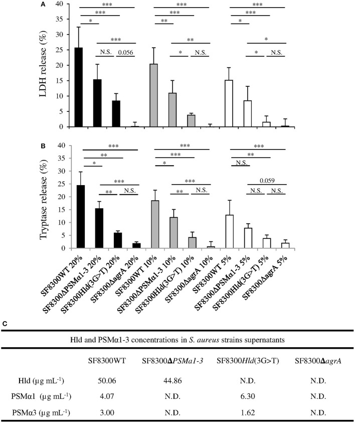
Figure 2.
Effect of SF8300WT, SF8300Δpsmα1-3, SF8300Hld(3G >T), and SF8300ΔagrA on human mast cells. HMC-1 cells were incubated with 20, 10, or 5% v/v S. aureus supernatants for 3 h at 37°C. (A) LDH and (B) tryptase release was measured in cell supernatants by Architect (Abbot®) and Immunocap (Phadia®), respectively. The percentage of mast cell lysis and mast cell degranulation was calculated as . The negative control and positive control were performed with medium and lysis buffer, respectively. The values represent the mean + SD of at least three independent experiments. (C) Hld, PSMα1, and PSMα3 concentrations in in each supernatant. To compare LDH and tryptase release by HMC-1 cells challenged with SF8300WT, SF8300Δpsmα1-3, SF8300Hld(3G > T), or SF8300ΔagrA supernatants, we performed t-test with Bonferroni correction followed by ANOVA. * p ≤ 0.05, ** p ≤ 0.01, *** p ≤ 0.001. N.S., not significant; N.D., not detected (toxin concentrations were less than the detection limit of 0.1 μg/mL).

