
Figure 2.
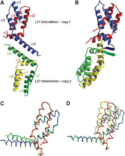
Structure of the L27PALS1N/L27PATJ heterodimer. (A) Overall structure of the two PALS1–PATJ L27 domain complexes present in the crystallographic asymmetric unit. Red/yellow: PALS1; blue/green: PATJ. (B) Ribbon diagram of the SAP97 L27 and the mLin2 L27N complex solved by NMR (Feng et al, 2004). Based on sequence alignment (Figure 1B) and domain classification, the SAP97 L27 domain is colored in blue and green, analogous to the PATJ L27 domain, whereas mLin2 is colored in red and yellow, analogous to the PALS1 L27N domain. Note the similarity in the heterodimer formation to that of the L27PALS1N/L27PATJ heterodimer, and the striking difference in the interface between the two heterodimers. (C) Overlay of the two heterodimers in the asymmetric unit based on Cα atoms belonging to Helix 1 and Helix 2 of the PATJ L27 domain. Red/yellow: PALS1; blue/green: PATJ. (D) Overlay of the two heterodimers in the asymmetric unit based on Cα atoms from Helix 3 of the PATJ L27 domain. All structural figures were generated with MOLSCRIPT (Kraulis, 1991) and RASTER3D (Merrit and Murphy, 1994).