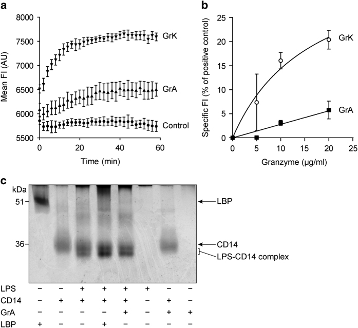
Figure 7.
GrA does not efficiently remove LPS from micelles and does not augment LPS-CD14 complex formation. (a) LPS-BODIPY-FL-FL, of which the fluorescent intensity (FI) increases upon removal from LPS micelles, was incubated for 90 min at 37 °C, after which granzymes (20 μg/ml) or extra PBS (LPS-BODIPY-FL control) were added. The FI was then measured for an additional 60 min. Results are depicted as mean±S.D. (n=3) and are representative of two independent experiments. Explanation of legends in figure: GrA=LPS-BODIPY-FL with GrA; GrK=LPS-BODIPY-FL with GrK, Control+LPS-BODIPY-FL alone. (b) LPS-BODIPY-FL was incubated with GrA or GrK and the mean FI was measured. Data are corrected for the FI of LPS-BODIPY-FL alone and depicted as the percentage of the FI of LPS-BODIPY-FL treated with 2% SDS. Data represent mean±S.D. (n=6). (c) LPS (2.5 μg) was incubated with human recombinant CD14 (0.5 μg) with or without LBP (0.5 μg) or GrA (0.5 μg) for 2 h at 37 °C. LPS-CD14 complex formation was analyzed by a band shift on native polyacrylamide gel electrophoresis (PAGE) followed by silver staining. Band intensities were quantified and showed that GrA did not augment LPS-CD14 complex formation.

