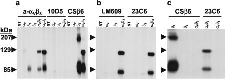
FIG. 1.
Analysis of soluble integrins secreted from transfected cells. COS-1 cells were transfected with soluble integrin subunits, as denoted in the figure, and labeled with [35S]methionine-cysteine between 24 and 72 h after transfection as described in Materials and Methods. The medium was collected, and the presence of soluble integrins or integrin subunits was determined by RIP, using the antibodies denoted in the figure followed by nonreducing SDS-PAGE and autoradiography. NT, nontransfected cells. The specificities of the antibodies were as follows: rabbit polyclonal anti-αVβ3 antibody (anti-αVβ3); monoclonal anti-αVβ6 antibody (10D5); monoclonal anti-β6 antibody (CSβ6); monoclonal anti-αVβ3 antibodies (LM609 and 23C6).

