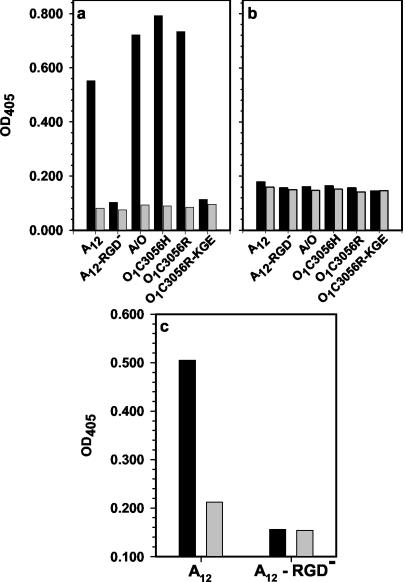
FIG. 2.
Analysis of binding of soluble αVβ3 and αVβ6 to FMDV. (a and b) Virus (1 μg) was captured on antibody-coated plastic wells as described in Materials and Methods. Culture medium from nontransfected HEK 293A cells (grey bars) or from transfected cells containing either soluble αVβ6 (a) or αVβ3 (b) (0.5 μg/well; black bars) was incubated with the virus, and binding of integrin was determined using either rabbit anti-αVβ3 (a) or MAb CSβ6 (b) as described in Materials and Methods. (c) Wells were coated with either medium from nontransfected cells (grey bars) or from transfected cells containing soluble αVβ3 (1 μg/ml; black bars) as described in Materials and Methods. Virus (1 μg) was incubated with the integrin, and the binding of virus was determined using an antiviral MAb.

