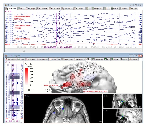
Figure 3.

HFO mapping of initial ictal activity at the orbitofrontal and mesial temporal ictal activity of a typical seizure. The EEG data on top depicts the time window where the HFO power was calculated. The bottom image on the left side displays the graphic bar power on each EEG channel and the right side represents the localization onto a 3D model based on the patient's own MRI.
