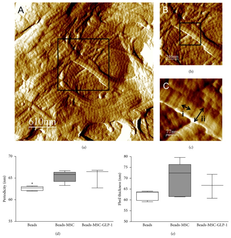
Figure 6.
Collagen fibril structures identified using AFM. (a) Fibre bundles from an untreated scar four weeks following MI. (b) A higher magnification of micrograph (a) reveals more details of the individual fibrils comprising the bundle, with D-periods clearly visible. (c) D-period length (i) and fibril width (ii) are indicated by arrows. There were a significantly increased periodicity (d), P < 0.05, and a trend towards increased fibril thickness (e) in MSC treated groups four weeks after MI.

