FIG.1.
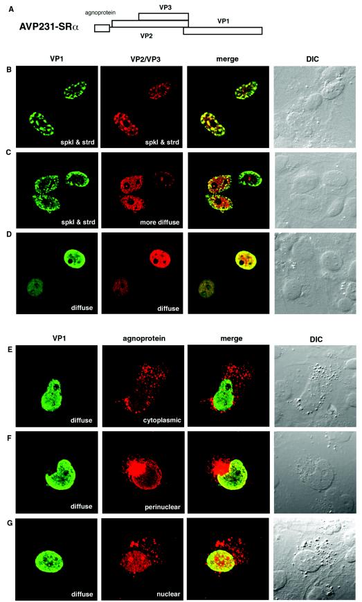
Localization of VP1, VP2/VP3, and agnoprotein in COS-7 cells transfected with AVP231-SRα. (A) Schematic illustration of AVP231-SRα expression vector. AVP231-SRα encodes a polycistronic fragment for agnoprotein, VP1, VP2, and VP3. (B to G) Immunocytochemistry of transfected cells. COS-7 cells transfected with AVP231-SRα were fixed at 3 days posttransfection. VP1 was visualized with Alexa fluor 488 (green), and VP2/VP3 or agnoprotein was visualized with Alexa fluor 568 (red). (B to D) Double staining of VP1 and VP2/VP3. The intranuclear distribution of VP1 and VP2/VP3 was variable. Both VP1 and VP2/VP3 show speckled staining patterns in panel B, there is a speckled staining of VP1 and a more diffuse staining of VP2/VP3 in panel C, and both show diffuse staining patterns in panel D. (E to G) Double staining of VP1 and agnoprotein. Agnoprotein shows a cytoplasmic localization pattern in panel E, a perinuclear inclusion in panel F, and a predominantly nuclear localization in panel G. spkl & strd, speckled and stranded staining patterns.

