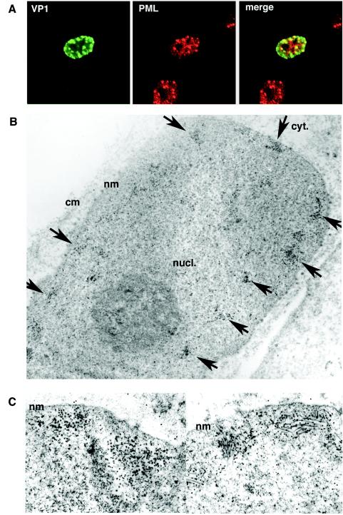
FIG. 9.
Capsid proteins accumulate at ND10 for assembly into virus-like particles. (A) Localization of VP1 at ND10. Cells transfected with AVP231-SRα were double stained for VP1 and PML, a component of ND10. VP1 was visualized with Alexa fluor 488 (green), and PML was visualized with Alexa fluor 568 (red). The two proteins were colocalized, showing speckled staining patterns at the inner periphery of the nucleus. (B) Immunoelectron microscopy of cells transfected with AVP231-SRα by use of an antibody against VP1. Colloidal gold spheres (15-nm diameter), indicating VP1 molecules, were clustered at multiple locations along the inner periphery of the nucleus (arrows). (C) Assembling virus-like particles with both spherical and filamentous structures were seen in association with clusters of colloidal gold. nucl., nucleus; cyt., cytoplasm; nm, nuclear membrane; cm, cellular membrane.

