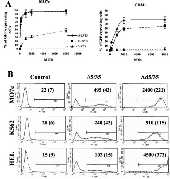
FIG. 2.
Transduction efficiency of different cell lines varies significantly for Ad vectors with different genome sizes. (A) Immortalized MO7e or primary human CD34-positive cells were infected at the indicated MOIs (virus particles per cell), and the percentage of GFP-expressing cells was analyzed 24 h later by flow cytometry (n= 6). (B) Immortalized human MO7e, K562, or HEL cells were infected at an MOI of 200 virus particles per cell with Δ5/35 or Ad5/35 vectors. At 24 h postinfection, the transduction efficiency was analyzed by flow cytometry. Mean GFP fluorescence intensities and standard deviations (in parentheses) are indicated on the corresponding histograms (n = 4). In the control settings (Control) cells were incubated with virus dilution buffer only.

