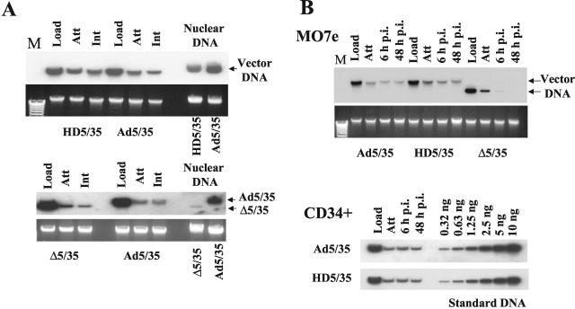
FIG. 5.
Delivery of viral DNA into the nucleus and its stability in infected cells over time. (A) MO7e cells were infected with the indicated Ad vectors for 8 h. Then, nuclei were extracted and nuclear DNA was purified for Southern blot analysis. To ensure that equivalent virus doses were used for initial cell infection and that all vectors were able to attach to and internalize into cells with similar efficiencies, at the corresponding time points duplicates of infected cells were lysed, and total cellular DNA was extracted and used for Southern blot analysis. The lower panel shows the agarose gel before Southern blotting to demonstrate equivalent DNA loads. The membrane was hybridized with a GFP gene-specific 32P-labeled probe, and vector DNA was visualized by autoradiography. Corresponding vector DNA bands are indicated by arrows. Note that although the Δ5/35 and Ad5/35 vectors were able to attach to and internalize into cells with similar efficiencies, the amount of vector DNA associated with nuclei was dramatically lower for Δ5/35 compared to Ad5/35. (B) MO7e or human primary CD34-positive cells were infected with the indicated Ad vectors as described in Materials and Methods. At the indicated time points, cells were lysed and total cellular DNA was purified for Southern blot analysis. Note that HD5/35 vector is maintained in infected cells as efficiently as Ad5/35 vector. However, Δ5/35 vector DNA is rapidly removed from the infected cells and is not detectable at 48 h post infection by Southern blotting. Serial dilutions of Ad5/35 plasmid DNA of known concentration was applied on the agarose gel as standard DNA. Att, attachment; Int, internalization; Load, the amount of virus vector used for cell infection; M, molecular weight marker.

