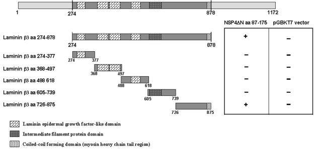
FIG. 4.
Binding domain analysis of laminin-β3 with NSP4ΔN. Laminin-β3 insert of clone D19 (aa 274 to 875) was fragmented into five subclones by PCR. Bars represent truncated laminin-β3 inserts fused to the GAL4 activation domain. Amino acid positions are indicated. Yeast strain AH109 was cotransformed with the laminin-β3 clones and pGBKT7-NSP4ΔN. The interaction was assessed by checking for blue colonies in the presence of X-α-Gal. Functional and structural domains of laminin-β3 are presented in boxes.

