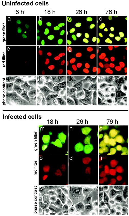
FIG.1.
Cytoplasmic fluorescence in poliovirus-infected HeLa cells expressing the Timer-NA-NLS fusion was due to the efflux of the presynthesized nuclear protein. In control uninfected Timer-transfected cells (a to l), the newly synthesized protein was accumulated in the nuclei and emitted fluorescence, which shifted with time from green to yellow when inspected with the green filter (a to d) and gradually became visible and increased in intensity when inspected with the red filter (e to h). In poliovirus-infected cells at 4 h p.i. (m to u), fluorescence could be seen not only in the nuclei but also in the cytoplasm, and the colors of nuclear and cytoplasmic fluorescence in a given cell coincided. The exposures were 10, 5, and 2 s for the pictures taken at 6, 18 to 26, and 76 h posttransfection, respectively, due to a marked difference in the fluorescence levels.

