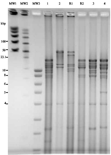
FIG. 6.
Cleavage patterns of the DNAs of BoHV-1.2 (lane 1), BoHV-5 (lane 2), recombinant 1 (GFP) (R1 from BoHV-1.2/ΔgC-GFP-ΔgI-RFP/BoHV-5 coinfection), recombinant 2 (RFP) (R2 from BoHV-1.2/ΔgC-GFP-ΔgI-RFP/BoHV-5 coinfection), BoHV-1.2/ΔgC-GFP (lane 3), and BoHV-1.2/ΔgI-RFP (lane 4). The DNAs were digested with HindIII and fragments were separated by PFGE. Fragments were visualized by ethidium bromide. Positions of molecular weight markers (MW) are indicated.

