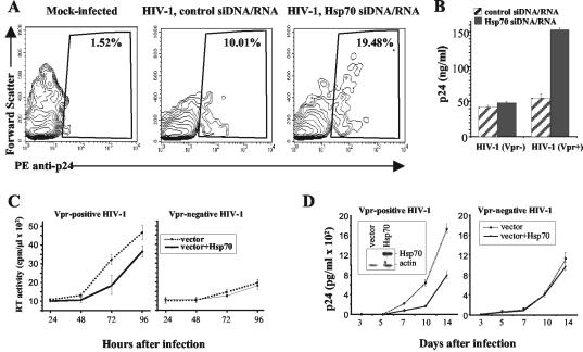
FIG. 3.
Analysis of Hsp70 effects on HIV-1 replication. (A) MAGI cells were transfected with control or Hsp70 siDNA/RNA. Twenty-four hours after transfection, cells were infected with HIV-1 or mock infected. HIV-1 replication was analyzed 48 h after infection by staining intracellular p24 essentially as described previously (32). The percentage of p24-positive cells is shown. For mock-infected cells, only results with Hsp70 siDNA/RNA-transfected cells are presented. (B) The experiment was performed as in panel A, except that cells were infected with Vpr-positive (Vpr+) or Vpr-negative (Vpr−) HIV-1 and extracellular p24 was measured by ELISA. Results show mean ± standard deviation of three independent wells. (C) Triplicate cultures of MAGI cells, transfected with either an Hsp70-expressing (vector+Hsp70) or an empty vector, were infected with Vpr-positive or Vpr-negative HIV-1. Virus replication was measured at indicated times after infection by reverse transcriptase activity in culture supernatants. Results are presented as means ± standard deviation. (D) Triplicate cultures of H9 cells stably transfected with Hsp70-expressing (vector+Hsp70) or an empty vector were infected with Vpr-positive or Vpr-negative HIV-1. p24 in culture supernatants was measured at indicated time points after infection by ELISA. Results are presented as the mean ± standard deviation. The inset in the left panel shows Western blot analysis of Hsp70 and β-actin proteins in transfected H9 cultures.

