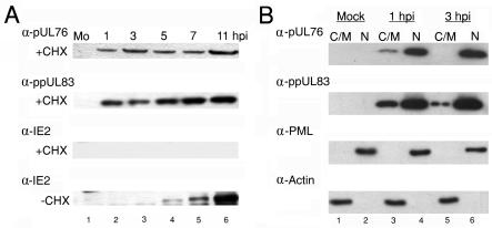
FIG. 2.
(A) Detection of pUL76 in HCMV-infected cells when protein synthesis was inhibited. For 1 h before infection with HCMV and continuing during the period of infection, cells were incubated in the presence of the protein synthesis inhibitor cycloheximide (+CHX; 100 μg/ml). Cell lysates harvested at the indicated times (hours postinfection) were subjected to Western blot analyses by incubating them with anti-pUL76, anti-ppUL83, and anti-IE2 antibodies (panels from the top down, respectively). As controls, infected cultures were grown in the absence of cycloheximide (−CHX). Cell lysates prepared at the indicated times (hours postinfection) were analyzed by Western blotting with an anti-IE2 antibody (last panel). Lanes 1 to 6, Western blotting of treated cell lysates harvested from mock-infected (Mo) cells and 1, 3, 5, 7, and 11 h postinfection with HCMV, respectively. (B) Western blot analyses of pUL76 and ppUL83 prepared from HCMV-infected cell subcellular fractions. The cytoplasmic/membrane (C/M) fractions of mock-infected (lane 1) and HCMV-infected HEL cells at 1 and 3 h postinfection (lanes 3 and 5, respectively) as well as nuclear fractions (N) of mock-infected (lane 2) and HCMV-infected HEL cells at 1 and 3 h postinfection (lanes 4 and 6, respectively) were resolved by SDS-10% PAGE. Proteins on Western blots were detected with anti-pUL76 and anti-ppUL83 antibodies. As controls, the membranes were subsequently stripped and analyzed with anti-PML protein and antiactin antibodies.

