FIG. 6.
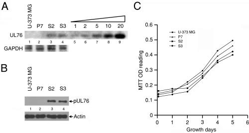
Analyses of U-373 MG cells and cells stably transfected with the cloning vector (P7 cells) or the UL76 gene (S2 and S3 cells). (A) Genomic Southern blotting for quantification of the UL76 gene. Lanes: 1, U-373 MG cells; 2, P7 cells transfected with the cloning vector pBK-CMV; 3 and 4, S2 and S3 cells, respectively, transfected with pUL76-CMV digested with SmaI. In lanes 5 to 9, 1, 2, 5, 10, and 20 pg, respectively, of purified UL76 DNA (SmaI-SmaI fragment, nucleotides 110228 to 111437) was loaded on the gel to be used as standards for relative quantification of the UL76 signal. The transfected UL76 gene was detected by probing with a riboprobe generated from pRB-UL76. As an internal control, the membrane was stripped and probed with the GAPDH gene. (B) Cell lysates prepared from the cells were immunoblotted with an anti-pUL76 antibody. As a control, the membrane was stripped and analyzed with an antiactin antibody. (C) Growth curve for various cells. A thousand cells were seeded per well in 96-well dishes and incubated until harvested at the indicated times. Cell growth was monitored by an MTT reduction assay as described in Materials and Methods. OD, optical density.

