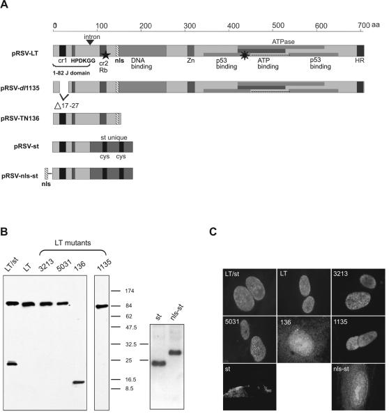
FIG. 1.
Schematic representation of the domain structure, expression, and subcellular localization of SV40 proteins encoded by related plasmids. (A) Schematic representation of the domain structure of SV40 proteins. pRSV-LT encodes the 708-aa-long SV40 LT, in which the depiction of the domain structure of LT has been adapted from Srinivasan et al. (43). The first 1 to 82 aa (1-82 J domain) in the N terminus represent the J domain containing cr1 and HPDKGG motifs. cr2 is a conserved motif allowing LT to bind pRb. The positions of the DNA binding domains, zinc finger motif (Zn), and sequences required for ATPase activity and p53 association are shown. HR, host range domain required for viron assembly. pRSV-dl1135 synthesizes a LT missing the cr1 motif due to a deletion (Δ17-27). PRSV-TN136 expresses the 136 N-terminal aa of LT containing the J domain, pRb binding domain, and NLS. pRSV-st directs the synthesis of wild-type SV40 st, which contains the first 1 to 82 aa of the J domain identical to that of LT and its unique C terminus. pRSV-nls-st expresses an st N terminally fused to an engineered NLS. The eight-pointed star indicates LT mutant pRSV-5031 lacking p53 binding function because of multiple mutations (D402N, V404M, and V423M) in the ATPase/p53 binding domain. ★, the position of two amino acid substitutions (E107K and E108K) within cr2 leading to the mutant pRSV-3213, which is defective in pRb binding ability; ▾, 448-bp intron (bp 4918 to 4571) in the SV40 LT early region following a 246-bp sequence (5163 to 4918) that is shared in both LT and st and encodes the identical 1 to 82 aa of the J domain (3). Plasmid pRSV-SV40(LT/st) encodes two early region products, both wild-type LT and st. (B) Expression of SV40 proteins. Two days after DOTAP transfection, Saos-2 cells were lysed and subjected to Western blot analysis. (C) Subcellular localization of SV40 proteins. Five days after DOTAP transfection, Saos-2 cells were fixed for immunofluorescence microscopy. For both Western blot analysis and immunofluorescence assay, LT-mutated 1135 protein was detected by using antibody KT3 raised specifically against the C-terminal 11 aa of LT, whereas the other SV40 proteins were detected by using antibody 419, which is reactive with the first 82 N-terminal aa shared by both LT and st.

