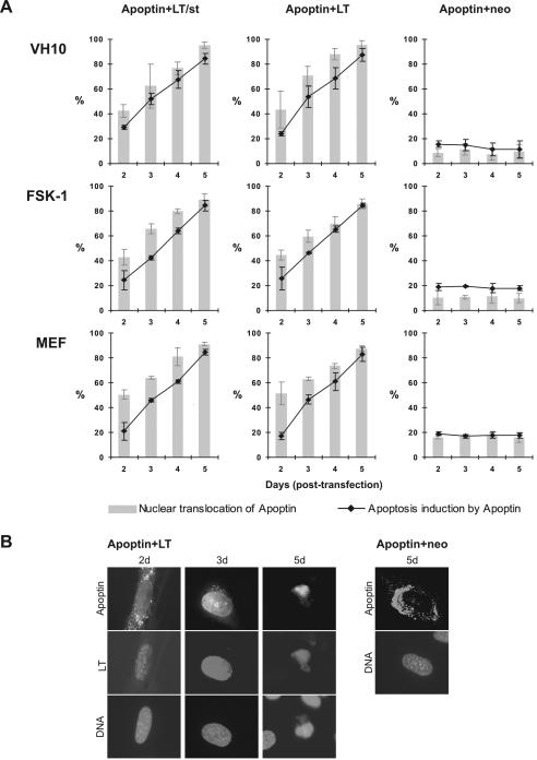
FIG. 2.
Activation of apoptin by the SV40 early region gene. Three different cell types (primary human diploid fibroblasts [VH10], human epidermal keratinocytes [FSK-1], and primary MEFs) were cotransfected with the apoptin-encoding plasmid pCMV-VP3 plus the plasmid pRSV-SV40(LT/st) encoding both LT and st, pRSV-LT expressing LT only, or empty vector pCMV-neo as a negative control. The expression and localization of apoptin was determined by apoptin antibody α-VP3-C, and SV40 early region products (LT/st or LT alone) were probed with antibody 419. DAPI staining was used to determine the nuclear morphology of cells indicative of apoptosis. (A) Transfected cells were analyzed at 2, 3, 4, and 5 days posttransfection by immunofluorescence microscopy. Three independent experiments were performed. Mean values are presented with standard deviation bars. Gray bars indicate the percentage of cells in which apoptin is mainly nuclear; black lines indicate the percentage of death. (B) Representative immunofluorescent images of VH10 cells showing apoptin's nuclear translocation (left and middle panels) and apoptosis induction (right panel) in the presence of LT and apoptin's primarily cytoplasmic localization in the negative control (Apoptin+neo). DNA was stained with DAPI (bottom of each panel) to show the fate of the cells with apoptin plus LT or with apoptin plus the empty vector pCMV-neo.

