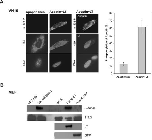
FIG. 5.
Activation of apoptin kinase by SV40 LT in normal cells both in vivo and in vitro. (A) In vivo phosphorylation of apoptin in VH10 cells. VH10 cells were comicroinjected with pCMV-VP3 and pRSV-LT and with pCMV-VP3 and pCMV-neo as negative control. 6 h postmicroinjection, coinjected cells were analyzed with the apoptin phosphospecific antibody α-108-P for phosphorylated apoptin (top), 111.3 for basal apoptin (middle images in the first and second panel), and 419 for coexpressed LT (middle image in the third panel). In addition, the bottom images of each panel show nuclear DNA staining with DAPI. The graph shows the percentage of phosphorylated apoptin as a ratio of α-108-P-positive cells to 111.3-positive-cells. (B) In vitro phosphorylation of apoptin in MEF cells. Using purified apoptin protein VP3-His as substrate, in vitro kinase assays were performed with MEF cells infected with LT-expressing retrovirus (Retrol-LT) or control GFP retrovirus (Retrol-GFP) 4 days postinfection, uninfected MEF (uninf.) as an additional negative control, and Saos-2 tumor cells as a positive control [Saos-2 (pos.)]. The blot was probed with antibody α-108-P for the phosphorylated apoptin, with 111.3 for the addition of apoptin-His protein, and with 419 and mouse α-GFP for the expression of SV40 LT and the control protein GFP, respectively.

