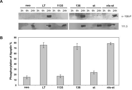
FIG. 6.
Apoptin kinase is activated by an N-terminal determinant of LT. The CD31-negative human primary fibroblasts were cotransfected using nucleofection transfection with pCMV-VP3 plus pRSV-LT, pRSV-dl1135, pRSV-136, pRSV-st, pRSV-nls-st, or pCMV-neo as a negative control and analyzed at the given times posttransfection by Western blot assay and parallel immunofluorescence microscopy. (A) Western blots showing the phosphorylated apoptin probed with α-180-P and the basal apoptin reprobed with 111.3. (B) Percentage of phosphorylation of apoptin at 24 h after transfection, scored as described in the legend to Fig. 5A. Two independent experiments were performed.

