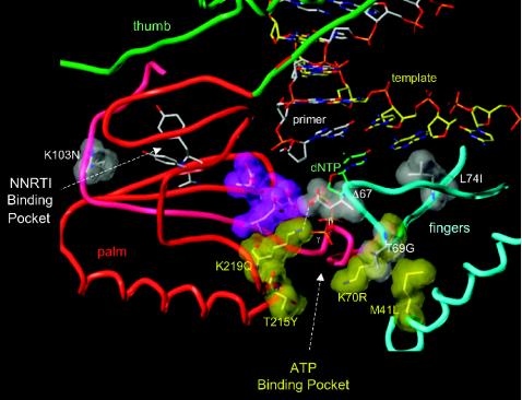
FIG. 1.
Model of the HIV-1 RT polymerase active site. The three aspartic acid residues (D110, D185, and D186) located at the active site are shown in purple. The subdomains of HIV-1 near the active site are color coded (palm, red; thumb, green; fingers, blue). A T/P is shown bound to the RT with the 3′ terminus of the primer in the P site. A dNTP is modeled in the N site, with the β and γ phosphates labeled. The nonnucleoside RT inhibitor (NNRTI) binding site is shown; the putative binding site for ATP located near the T215Y residue. The residues described in the text are shown in gray and yellow space-filling models; the amino acid residues in the figure are those normally found in wild-type HIV-1 RT. The labels indicate the amino acid substitutions found in the Δ67 complex.

