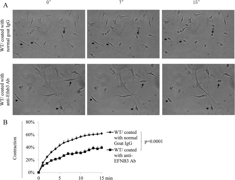
Figure 1. Crosslinking EFNB3 on VSMCs results in decreased contractility.
VSMCs from female WT mice were cultured in wells coated with goat anti-mouse EFNB3 Ab or control goat IgG (2 μg/ml during coating) for 4 days. The cells were then stimulated with PE (20 μmol/L), and their percentage contraction was recorded by microscopy. (A) Micrographs showing the contraction of WT VSMCs in the presence or absence of solid phase anti-EFNB3. Upper row: WT VSMCs cultured in wells coated with control goat IgG (20 μg/ml for coating). Lower row: WT VSMCs cultured in wells coated with goat anti-mouse EFNB3 Ab. The images were taken at 0, 7 and 15 min after PE stimulation. Arrows mark the same cells in each row at different time points, to show the shortening of the cells. (B) Reduced contractility of WT VSMCs cultured in the presence of solid phase anti-EFNB3 Ab. VSMCs from female WT mice were cultured in wells coated with normal goat IgG or goat anti-mouse EFNB3 Ab for 4 days. The cells were then stimulated with PE, and were imaged at one frame per min for 15 min. Means ± SD of the percentage contraction during a 15-min period are reported. The percentage contraction is calculated as follows. % contraction = length of cells at a given timepoint/length of the cells at time 0. The data were analyzed with one-way ANOVA, and p-value is indicated. The experiment in this figure was repeated three times, and representative data are shown.

