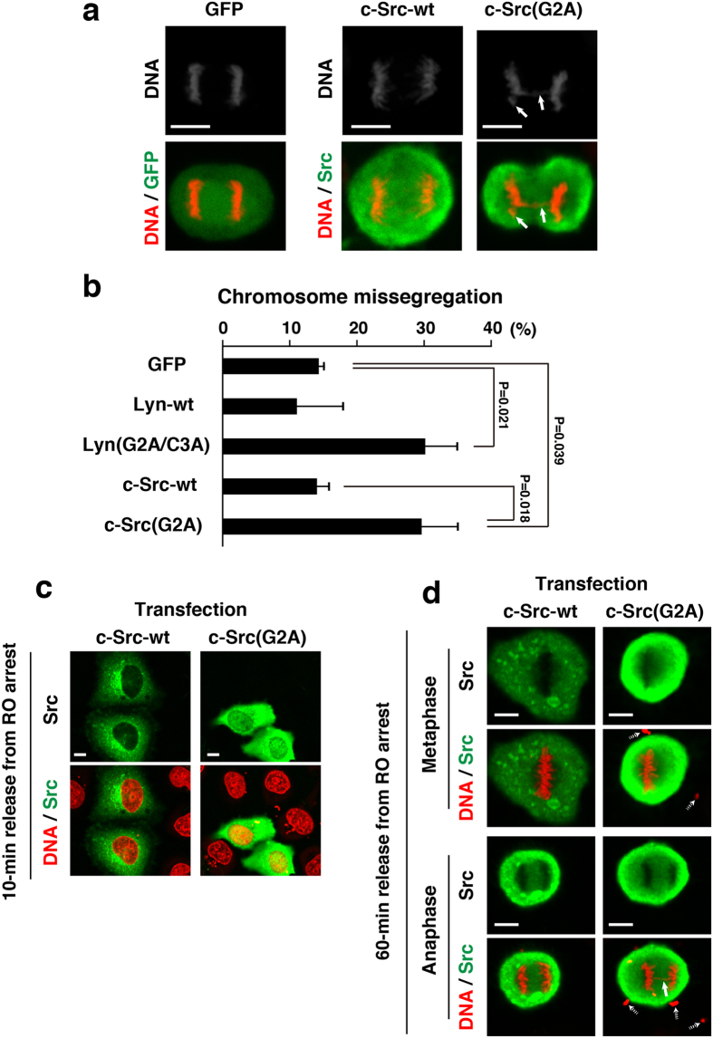
Figure 4. Protective role of Src myristoylation against chromosome missegregation.
(a) HeLa S3/TR cells transiently transfected with GFP, c-Src, or c-Src(G2A) were synchronized as shown in Fig. 3a. Bold arrows indicate a lagging chromosome and a chromosome bridge. (b) The cells exhibiting chromosome missegregationn (chromosome bridging and lagging) were quantitated (>62 cells). Values are means ± S.D. from more than three independent experiments, and the significant differences are calculated by Student’s t-test. (c,d) HeLa S3/TR cells transiently transfected with c-Src or c-Src(G2A) were synchronized as shown in Fig. 3a, and were released from RO-3306-treated arrest at the G2/M boundary for 10 min (early prophase; see Fig. 1) (c) and 60 min (metaphase and anaphase) (d). Cells were stained for Src proteins (green) and DNA (red). A bold arrow indicates a chromosome bridge. Dotted arrows show plasmid DNA polyplexes deposited extracellularly, which were added to cell cultures for transient transfection. Note that plasmid DNA polyplexes were often observed in transient transfection cultures but not inducibly expression cultures.

