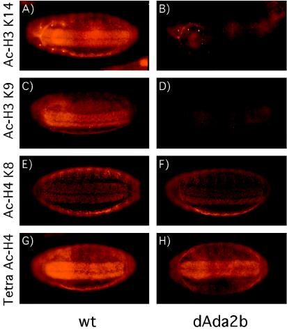
FIG. 3.
Reduced histone H3 acetylation in dAda2b mutant embryos. Antibodies that recognize different acetylated forms of histone H3 and histone H4 were used to stain Drosophila embryos. All four antibodies show strongest signals in the CNS and epidermis. Ventral views of stage 16 embryos are shown, with anterior to the left. Homozygous dAda2b mutant embryos were identified by the absence of Ubx-lacZ expression, which is present in embryos containing a balancer chromosome. (A and B) Wild-type (wt) (A) and dAda2b mutant (B) stage 16.1 embryos were stained with an antibody recognizing histone H3 acetylated at lysine 14. A substantial reduction in staining intensity was found in dAda2b mutant embryos. (C and D) wt (C) and dAda2b mutant (D) stage 16.1 embryos incubated with an antibody directed against acetylated histone H3 lysine 9. The staining intensity is diminished in dAda2b mutant embryos compared to wt embryos. (E and F) wt (E) and mutant (F) stage 16.2 embryos stained with an acetylated histone H4 lysine 8 antibody. The staining pattern and intensity are indistinguishable between wt and dAda2b mutant embryos. (G and H) Stage 16.2 wt (G) and dAda2b mutant (H) embryos incubated with a tetra-acetylated histone H4 antibody. Comparable staining intensities were observed in wt and mutant embryos.

