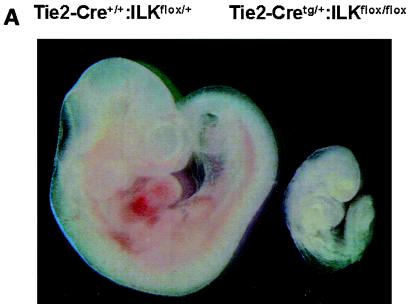
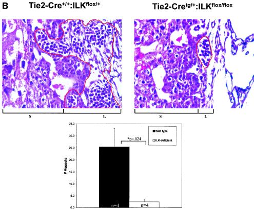
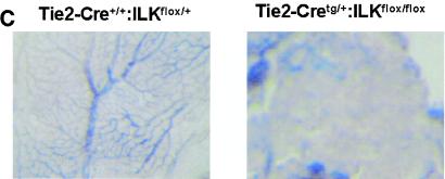
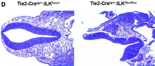
FIG. 1.
Endothelium-specific deletion of ILK in mice. Embryos, placentae, and yolk sac were harvested at E9.5 to E10.5. (A) Images of whole embryos of Tie2-Cre+/+:ILKflox/+ and Tie2-Cretg/+:ILKflox/flox genotypes. (B) Hematoxylin and eosin-stained section from placentae of Tie2-Cre+/+:ILKflox/+ and Tie2-Cretg/+:ILKflox/flox embryos. Chorionic/labyrinthine vessels (indicated by red outline), filled with nucleated red cells generated in the embryos, were counted from control (15.3 ± 7.6) and mutant (2.5 ± 0.0.87) placentae (n = 4; P = 0.024), below. S, spongiotrophoblast; L, labyrinth. (C) Whole-mount stained yolk sacs, using anti-PECAM antibody. (D) Hematoxylin and eosin-stained embryo sections from head region. Vessels are indicated with asterisks.


