FIG. 4.
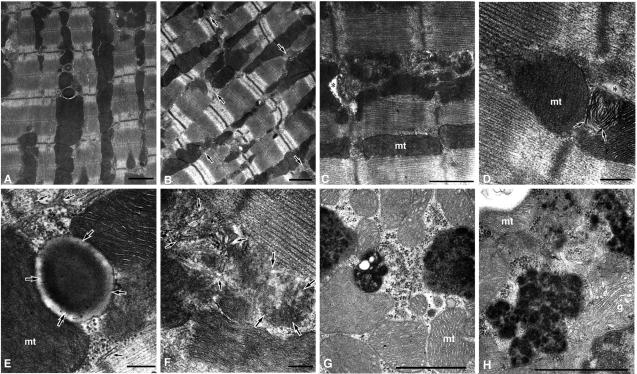
Transmission electron microscopic analysis of cardiomyocytes. The pictures in panels A and E are from a control C57BL/6 mouse (15 weeks old). The pictures in panels B and D are from mutant transgenic mouse line F without apparent cardiomegaly (13 weeks old), and the pictures in panels C, F, G, and H are from mutant transgenic mouse line B with apparent cardiomegaly (15 weeks old). All the mice were Cre gene positive. (A) Myocardium from the control. (B) Myocardium from mutant transgenic mouse F. Though the arrangement of myofibrils and the banding patterns of myofilaments are intact, the structures around the transverse tubules are aberrant (arrows). (C) Proliferation of the sarcoplasmic reticulum is evident, and the transverse tubule lumen is significantly narrowed (asterisk) in the mutant B. mt, mitochondrion. (D) Lamellated membrane structures (arrows) are found to be apposed to the narrowed T-tubule (asterisk) in the mutant F. (E) Higher magnification of a transverse tubule (large arrows) and surrounding structures, such as the sarcoplasmic reticulum (small arrows) and mitochondria, in the control. (F) The aggregation of degenerate membrane structures (large arrows) is found to be apposed to the proliferated sarcoplasmic reticulum (small arrows) in mutant B. (G and H) Electron-dense structures accumulated in the mutant cardiomyocytes were associated with polyribosomes. g, Golgi complex. Bars: panels A to C, G, and H, 1 μm; panels D to F, 200 nm.

