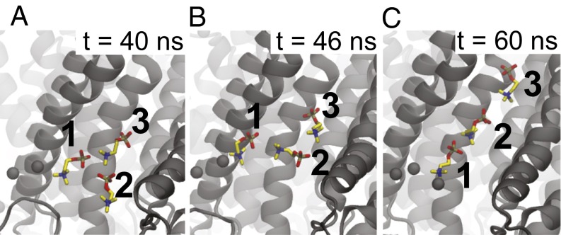
Fig. 3.
Lipid penetration into the groove occurs via a dipole-stacking mechanism. A–C are sequential MD snapshots during the lipid-flopping event. The negative phosphate groups (gold) interact with the positive choline groups (blue) of neighboring lipids. Lipid 2 inserts between lipids 1 and 3 and then stretches out to push lipid 3 far into the groove on the way to the extracellular space over the course of 20 ns. For clarity, only the phosphatidylcholine group is shown. The phosphorus atoms of all other lipids in the cytoplasmic leaflet are shown as black spheres.

