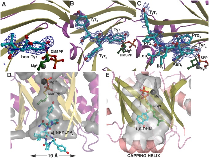
Fig. 3.
(A–C) Cocrystal structure of PagF and substrate DMSPP with boc-Tyr (A), Tyr-Tyr-Tyr (B), and cyclic[INPYLYP] (C). A simulated annealing omit (|Fobs − Fcalc|) difference Fourier electron density map is superimposed at 2.4σ (blue). The requisite magnesium ion is shown as a gray sphere. (D) Cutaway diagram showing the volume of the PagF active site viewed parallel to the orientation of the prenyl donor DMSPP and the acceptor cyclic[INPYLYP]. The dimensions of the base of the active site roughly correlate with the size of the natural substrate. (E) Cutaway diagram of the small-molecule prenyltransferase NphB with donor GSPP and acceptor 1,6-dihydroxynapthalene (1,6-DHN) showing a completely occluded active site chamber.

