Fig. 1.
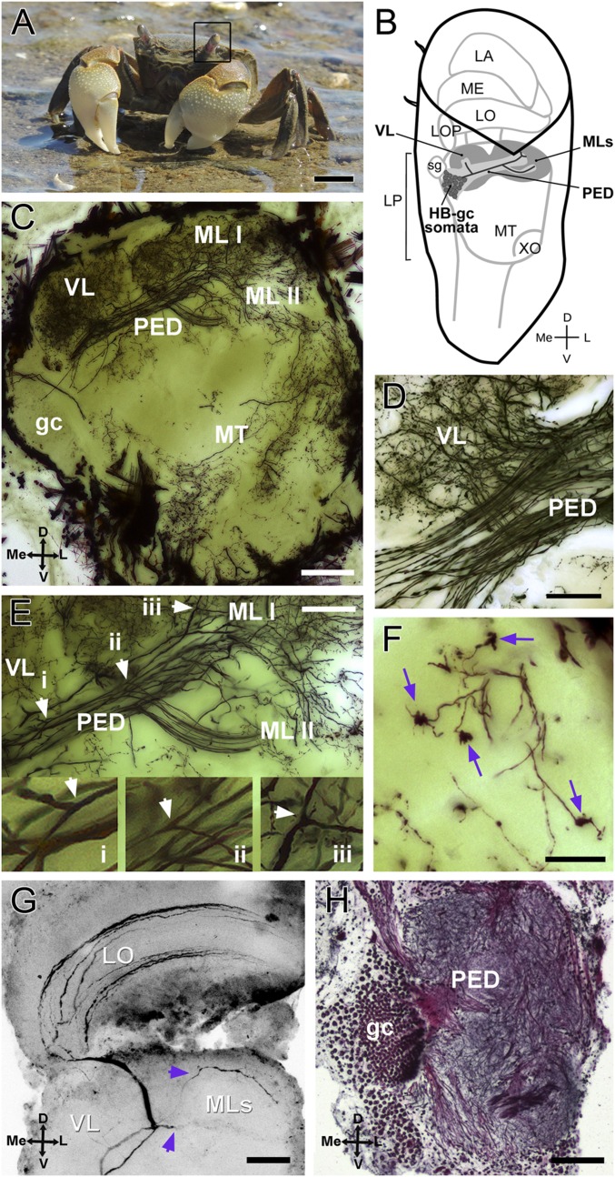
Crab N. granulata and the HB. (A) N. granulata crab. The black frame surrounds the left eyestalk. (B) Scheme showing the position and shape of the neuropils within the eyestalk. The HB is part of the lateral protocerebrum and is drawn in gray. Complete gc and some somata of the HB are drawn. (C–F) Golgi impregnation of the optic lobes (50-μm sections). (C) Overall structure of the HB seems mainly formed by intrinsic cells derived from the HB-gc cluster. (D) Detail of the bifurcation site that originates the vertical lobe. (E) Detail of the bifurcation sites that give rise to the vertical lobe and to both medial lobes. (Insets) Arrowheads show three amplified bifurcations (i, ii, and iii) in the same neuron. (F) Claw-like specializations are present in the medial lobes (arrows). (G) Intracellularly stained BLG2 neuron showing two branches that invade the two medial lobes of the HB (arrowheads). (H) Bodian-stained section (12 μm) of the lateral protocerebrum showing gc and the pedunculus. D, dorsal; L, lateral; LA, lamina; LO, lobula; LOP, lobula plate; LP, lateral protocerebrum; Me, medial; ME, medulla; MT, medulla terminalis; PED, pedunculus; MLs, medial lobes; sg, sinus gland; V, ventral; VL, vertical lobe; XO, X-organ. (Scale bars: A, 1 cm; C, G, and H, 100 μm; D and E, 50 μm; F, 25 μm.)

