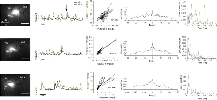
Fig. S2.
Spontaneous activity in the HBs. Examples of trained animals showing calcium transients in the absence of stimuli. (Left) Pictures of the registered area showing the AOI of the vertical lobe (VL) and medial lobes (MLs) from which the %ΔF/F was obtained. (Scale bar: 200 μm.) (Right) %DeltaF/F registered in the absence of explicit stimulation, correlation and cross-correlation, and power spectrum for the signals obtained in the vertical and medial lobes of the HBs. D, dorsal; Freq, frequency; L, lateral; Me, medial; V, ventral.

