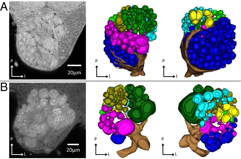
Fig. 4.
Antennal lobes of (A) female and (B) male clonal raider ants. From Left to Right: representative slices from confocal micrograph stacks used in reconstruction, dorsal (n-ventral) view of 3D reconstruction of glomeruli and antennal nerve, ventral (n-dorsal) view of 3D reconstruction of glomeruli and antennal nerve. P, posterior (n-rostral); M, medial; L, lateral; gold, T1; dark green, T2; purple, T3; cyan, T4; light green, T5; blue, T6; yellow, T7. Figs. S1 and S2 show complete 3D models of female and male antennal lobes, respectively.

