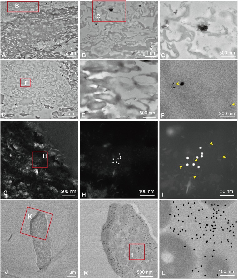
Fig. 4.
Immunogold labeling of fossil feathers (sample 201) compared with feathers of G. gallus. (A–C) Negative control of D–F, when the primary antiserum is omitted. (B) High-magnification image of the boxed area in A. (C) High-magnification image of the boxed area in B. (D–I) In situ nanogold immunohistochemistry of Eoconfuciusornis feathers exposed to anti-feather antibody. (E) Melanosomes from the same section of D and F; no nanogold particles were seen to specifically bind. (F) High-magnification image of the boxed area in D; arrowheads show binding of the nanogold beads. (G–I) High-angle annular dark-field (HADDF) images show binding of the nanogold beads from the same section of D–F. (H) High-magnification image of the boxed area in G. (I) High-magnification image of H; arrowheads show the filaments at the bead-binding regions. (J–L) In situ nanogold immunohistochemistry of G. gallus feathers exposed to anti-feather antibody. Nanogold particles specifically bind the keratinous tissues but do not bind the melanosomes, visualized in cross-section. (K) High-magnification image of the boxed area in J. (L) High-magnification image of the boxed area in K. (Scales are as indicated.)

