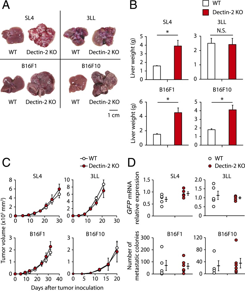
Fig. 1.
Selective contribution of Dectin-2 to the suppression of liver metastasis. (A and B) SL4 cells (2 × 10⁵ cells), 3LL cells (3 × 10⁵ cells), B16F1 cells (1 × 10⁶ cells), or B16F10 cells (2 × 10⁵ cells) were inoculated into the spleens of WT and Dectin-2 KO mice. Fourteen days later, the livers were observed macroscopically (A) and liver weights were measured (B). (Scale bar: 1 cm.) (C) SL4 cells (2 × 10⁵ cells), 3LL cells (5 × 10⁵ cells), B16F1 cells (5 × 10⁵ cells), or B16F10 cells (1 × 10⁵ cells) were inoculated s.c. into WT and Dectin-2 KO mice, and tumor volumes were measured every 3 or 4 d. (D) SL4-GFP cells (3 × 10⁵ cells), 3LL-GFP cells (1 × 10⁶ cells), B16F1 cells (1 × 10⁶ cells), or B16F10 cells (5 × 10⁵ cells) were inoculated i.v. into WT and Dectin-2 KO mice, and the metastatic levels of SL4-GFP and 3LL-GFP cells were evaluated by quantifying GFP mRNA in the lung on day 12. The numbers of B16F1 and B16F10 colonies in the lung were counted at 14 d after inoculation. Data are shown as mean ± SEM. *P < 0.05. N.S., not significant.

