Fig. 3.
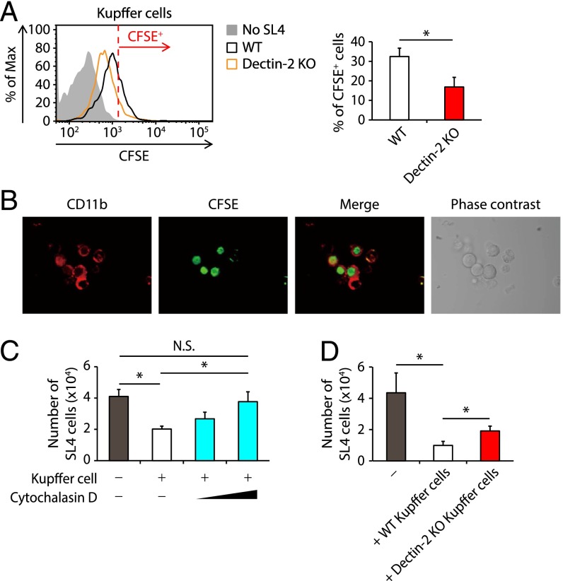
Dectin-2–dependent engulfment and clearance of cancer cells by Kupffer cells. (A) Kupffer cells (1 × 10⁵ cells) isolated from WT and Dectin-2 KO mice were cocultured with or without CFSE-labeled SL4 cells (0.25 × 10⁵ cells) for 2 h. The CFSE intensity in CD45+ CD11b+ F4/80+ cells was analyzed by flow cytometry. (Left) Representative histograms of CFSE level. The cells with a CFSE level exceeding the red line were identified as CFSE+ cells. (Right) Proportion of CFSE+ cells. (B) Kupffer cells (1 × 10⁵ cells) were cocultured with CFSE-labeled SL4 cells (0.25 × 10⁵ cells) and after 2 h, the cells were observed by confocal microscopy. (C) CFSE-labeled SL4 cells (0.25 × 10⁵ cells) were cultured in the presence or absence of Kupffer cells (1 × 10⁵ cells) pretreated with DMSO or cytochalasin D, and the number of PI− CD45− CFSE+ cells was determined at 24 h after culturing. (D) CFSE-labeled SL4 cells (0.25 × 10⁵ cells) were cultured in the presence or absence of Kupffer cells (1 × 10⁵ cells) derived from WT and Dectin-2 KO mice, and the number of PI− CD45− CFSE+ cells was determined at 24 h after culturing. Data are shown as mean ± SEM. *P < 0.05. N.S., not significant.

