Fig. S1.
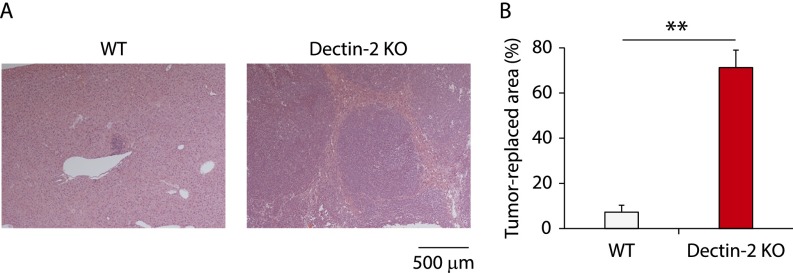
Histological analysis for Dectin-2–mediated suppression of liver metastasis. At 14 d after WT and Dectin-2 KO mice were inoculated intrasplenically with SL4 cells (2 × 10⁵ cells), liver sections were analyzed by H&E staining. Representative staining images (A) and tumor-replaced areas (B) are shown. (Scale bar: 500 μm.) Data are displayed as mean ± SEM. **P < 0.01.

