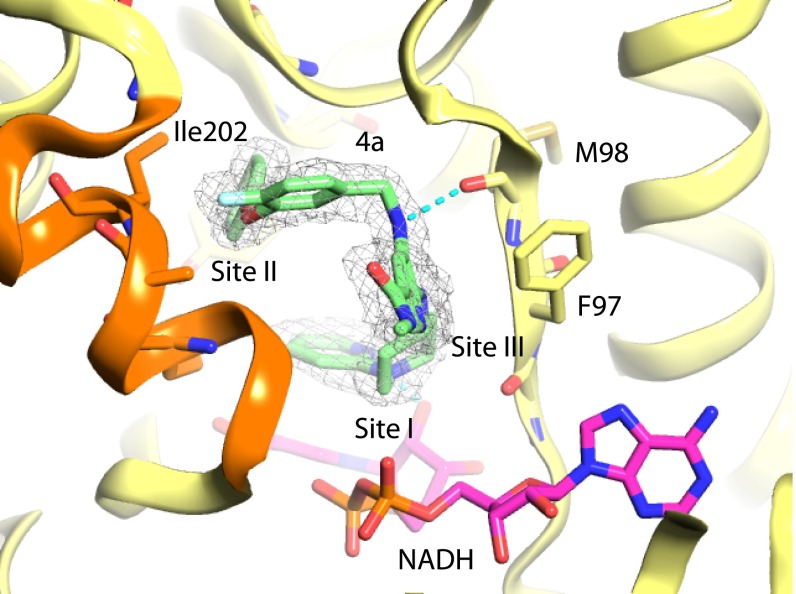
Fig. 3.
X-ray crystal structure of InhA:NADH in complex with compound 4a (PDB ID code 5G0U) from series 4. Carbon atoms for compounds are shown in green. The protein backbone cartoon is represented in yellow. Selected atoms for the InhA side chains including F97, M98, and Ile202 in the active site loop are shown as sticks. NADH is shown as sticks with magenta carbon atoms. Refined (2fo-fc) electron density contoured at 1.0 σ is represented as a wire mesh. Some atoms of the active-site covering loop represented in orange have been removed for clarity.

