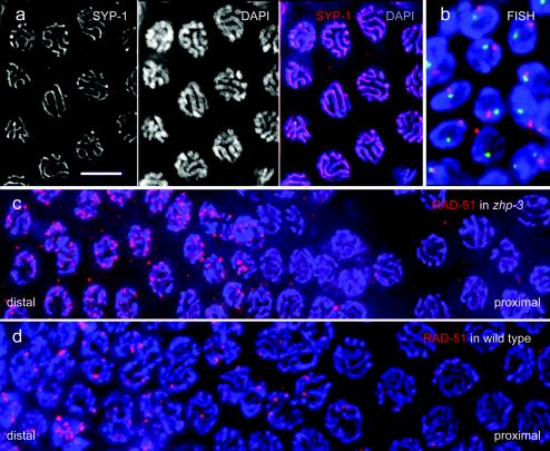
FIG. 4.
Pairing and localization of recombination protein in the mutant. (a) SYP-1 localizes to the gap between paired chromosomes as visualized by DAPI, as can be seen in the merged image. (b) FISH of the 5S rDNA (red) and a locus on chromosome I left arm (green) in pachytene nuclei of the zhp-3 mutant. The presence of a single FISH signal per nucleus for each of the two loci tested is evidence for homologous pairing. Sectors with late pachytene nuclei of gonads of (c) the zhp-3 mutant and (d) the wild type immunostained for the Rad-51 recombination protein (red). Note the larger number of Rad-51 foci and their sudden disappearance before the end of pachytene in the mutant (distal-proximal indicates the orientation of the sector shown within the gonad). Chromatin is stained blue by DAPI. Bar, 5 μm.

