FIG. 10.
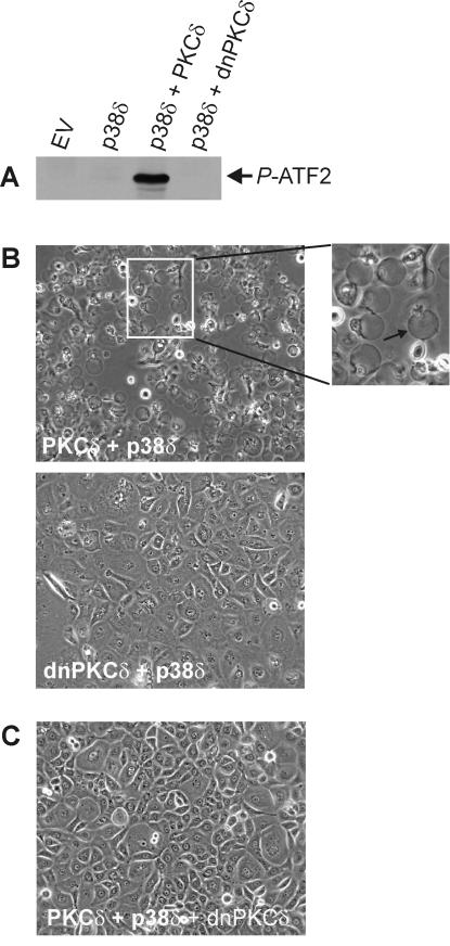
PKCδ kinase activity is required for p38δ activation and appearance of apoptotic morphology. (A) Keratinocytes were infected with empty virus (EV) (MOI = 30), PKCδ or dnPKCδ-encoding virus (MOI = 15) plus FLAG-p38δ virus (MOI = 15), or EV (MOI = 15) with p38δ virus (MOI = 15) for 48 h. Total cell extracts were prepared, FLAG-p38δ was immunoprecipitated by using anti-FLAG, and the activity of the immunoprecipitated p38δ was measured by using in vitro kinase with ATF2 as a substrate. The P-ATF2 level was measured by immunoblotting with anti-phospho-ATF2. The total p38δ level was found to be equal in each infection as measured by immunoblotting with anti-FLAG (not shown). An immunoblot of extracts prepared from PKCδ- and dnPKCδ-expressing cells reveals comparable levels of expression at approximately four times endogenous levels (not shown). (B) Keratinocytes were infected with PKCδ+p38δ or dnPKCδ+p38δ as described above and photographed after 48 h. The arrow in the enlarged panel indicates the apoptotic spheres. (C) Keratinocytes were triply infected with of PKCδ, p38δ, and dnPKCδ (MOI = 15 for each) and photographed after 48 h.

