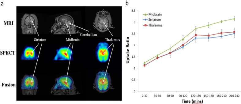
Figure 1.
(a) Representative images of MRI and [123I]-ADAM/SPECT in coronal (left column), sagittal (middle column) and horizontal (right column) views. (b) Uptake ratios of [123I]-ADAM in the midbrain, striatum and thalamus at different time points in normal monkeys. The data are expressed as the mean ± standard deviation (S.D.).

