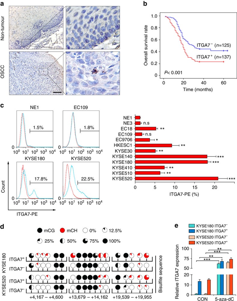
Figure 1. High frequency of ITGA7+ cells is significantly associated with poor outcome in OSCC.
(a) Representative IHC images show that ITGA7+ cells were scattered in OSCC tumour tissue in clinical specimen, but not in non-tumour tissue. Scale bar, 100 μm. (b) Kaplan–Meier survival analysis shows that OSCCs with high frequency of ITGA7+ cells (>0.6%, ITGA7+, n=137) had shorter survival time, compared with OSCCs with low frequency of ITGA7+ cells (≤0.6%, ITGA7−, n=125). (c) Percentage of ITGA7+ cells detected by FACS in immortalized esophageal epithelial and OSCC cell lines. The average percentage of ITGA7+ cells, the mean±s.d. of three independent detections, in different cell lines was depicted in the bar chart. (d) Detection of DNA methylation in the CG and CH context (H=A, C or T) by genomic bisulfite sequence. Non-CG methylation of ITGA7 preferred to occur in ITGA7+ cells isolated from KYSE180 and KYSE520. (e) qRT-PCR showed that the expression of ITGA7 was markedly increased after treated with 5-aza-2′-deoxycytidine (5-aza-dC, 50 μM) for 3 days. Statistics: (c,e) ANOVA with post hoc test. *P<0.05; **P<0.001; ***P<0.0001; n.s., P≥0.05.

