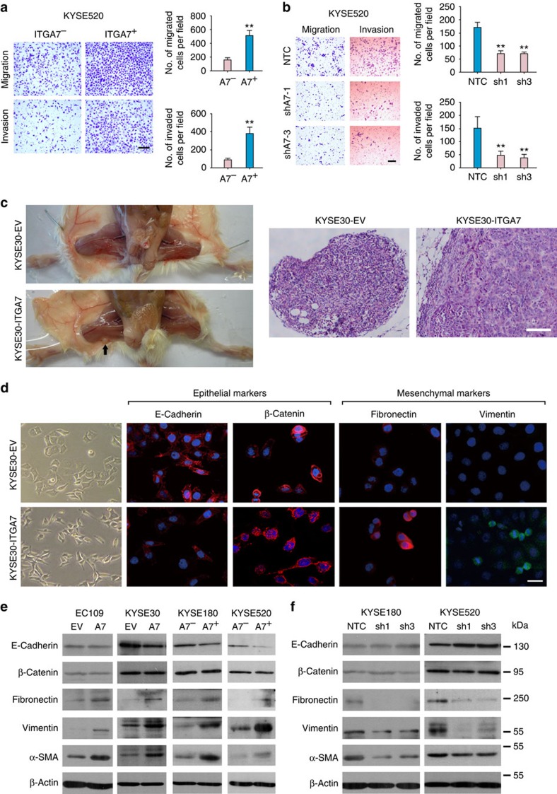
Figure 5. ITGA7 promotes metastasis by inducing EMT.
(a) Representatives and summary of migration and invasion assays showing that ITGA7+ cells exhibited enhanced migration and invasion abilities. The values expressed as the mean±s.d. of three independent experiments. Scale bar, 200 μm. (b) Representatives and summary of migration and invasion analyses showing that knockdown of ITGA7-suppressed cell motility of KYSE520, compared with their control cells. The values expressed as the mean±s.d. of three independent experiments. Scale bar, 200 μm. (c) Representatives of lymph node metastasis formed in NOD/SCID mice injected with ITGA7- or EV-transfected KYSE30 cells. Black arrow indicates the swollen popliteal lymph node. Lymph nodes invaded by tumour cells were confirmed by H&E staining (Scale bar, 100 μm). (d) Cell morphology of K30-ITGA7 and K30-EV cells. Epithelial markers (E-cadherin and β-catenin) and mesenchymal markers (fibronectin and vimentin) were detected by immunofluorescence staining (red: E-cadherin, β-catenin and fibronectin; green: vimentin; blue: nuclear DAPI staining; scale bar, 50 μm). (e,f) Expression of EMT markers in indicated cells was detected by western blot analysis. β-Actin was used as a loading control. Statistics: (a) Student t–test. (b) ANOVA with post hoc test. **P<0.001.

