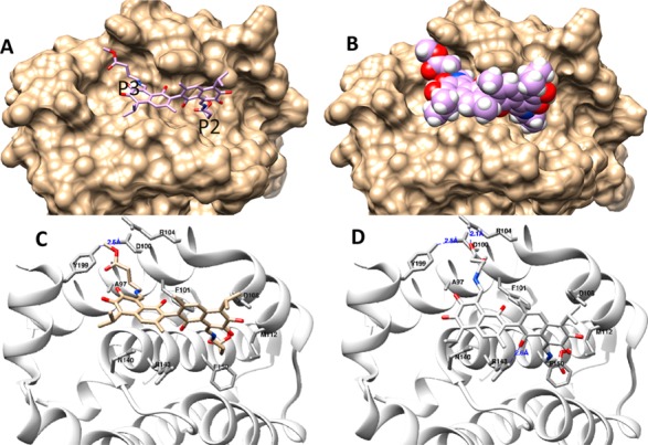
Figure 4.

(A,B) The interaction models of compound (−)-16 with Bcl-2. (A) The binding site and orientation of the compound (represented by compound (−)-16) in the hydrophobic groove P2 and P3 of Bcl-2. (B) The protein is rendered in a surface model, while the compound is rendered in a stick model and a van der Waals sphere (CPK) model. (C,D) Two possible poses were assessed in the molecular docking study, (C) with no ligand-protein hydrogen bonding interaction and (D) with a ligand–protein hydrogen bonding interaction; Bcl-2 is rendered as a cartoon, while the compound and the residues around it are rendered as sticks (enlarged pictures, see Supplemental Figure S11).
