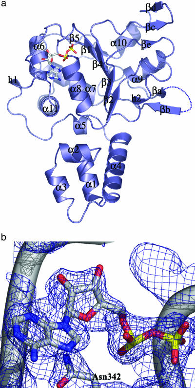
Fig. 1.
The Rep40–ADP complex. (a) The AAV2 Rep40 molecule (slate), complexed to ADP at 2.1 Å. The nucleotide sits in an unexpectedly open binding site formed by the P-loop residues and the NB-loop. The β-hairpin 1 loop (βa–βb) is disordered, with electron density for five consecutive residues, 402–407, missing. There are two secondary elements: a βe strand that is part of a three-stranded β-sheet together with strands βc–βd and a 310-helix h2 that is between β2 and β3. (b) A 2Fo – Fc simulated annealing omit map showing the electron density for the bound ADP at 1.5 σ.

