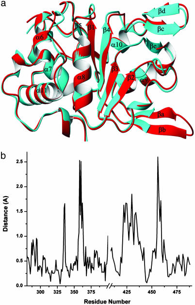
Fig. 3.
Comparison of the Rep40 apo and ADP-bound structures. (a) Superposition of the AAV2 Rep40 apo (red) and ADP-bound (cyan) structures. Small differences are observed in response to ADP binding. (b) Plot of the average difference distance (Å) versus residue number for the two superimposed molecules. Four regions of differences are seen: the P-loop between β1 and α8, the quasihelical loop connecting β2 and β3, the β-hairpin 2 (βc–βd) loop, and the NB-loop. Differences in the β-hairpin 1 (βa–βb) loop may be caused by the lack of crystal contacts in the ADP-bound form of the protein.

