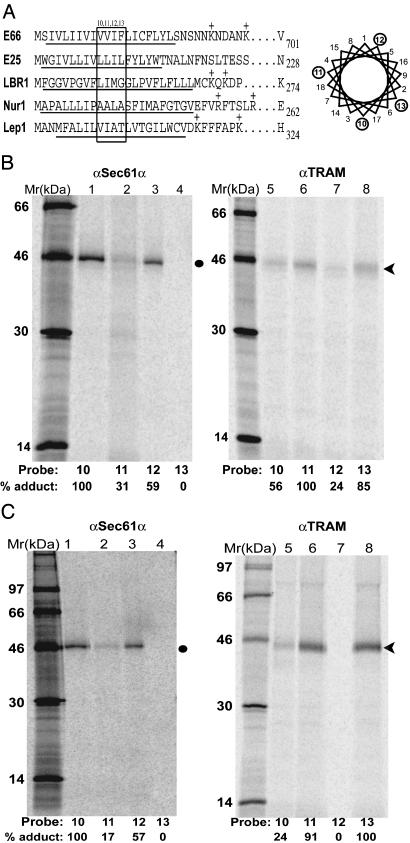
Fig. 1.
Photocrosslinking of viral INM-directed TMSs to translocon proteins. (A) The N-terminal sequences of E66 and E25 are shown with the TMS underlined, as are the N-terminal sequences of the constructs containing the first TMS of LBR (LBR1), nurim (Nur1), and Lep (Lep1). In each case, an amber codon was substituted for the codon shown at position 10, 11, 12, or 13 (boxed in figure) to position the photoreactive probes at a single nascent chain location in each sample. The probes extend from different sides of the TMS α-helix surface as shown in the helical wheel representation. (B) Integration intermediates containing 70-residue E66-A10, E66-A11, E66-A12, or E66-A13 nascent chains were photolyzed, immunoprecipitated with affinity-purified antibodies to Sec61α (lanes 1–4) or TRAM (lanes 5–8), and analyzed by SDS/PAGE. Photoadducts to Sec61α and TRAM are indicated by the closed circle and arrowhead, respectively. The relative extent of photoadduct formation was quantified by comparing each photoadduct band intensity with that of the most intense photoadduct band in the gel (assigned 100%). (C) Integration intermediates containing 70-residue E25-A10, E25-A11, E25-A12, or E25-A13 nascent chains were photolyzed and analyzed as in B.

