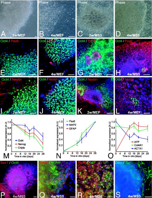
Fig. 1.
Stromal feeder-induced neural differentiation of hES cells. (A–D) Representative phase contrast images of hES cells (line H1) cocultured for 2 or 4 weeks on MEF (A and B) or MS5 (C and D). (E–H) Pax6 and Oct4 expression in hES cells differentiated on MEF or MS5. (I–L) Oct4 and nestin expression in hES cells on MEF or MS5. (M–O) Semiquantitative RT-PCR analysis for genes characteristic of undifferentiated ES cells (M); neural lineage (N); and non-neural, endodermal, and mesodermal differentiation (O). Data are presented as normalized values (see Materials and Methods) and derived from three independent experiments, line H1, days 0–28. GFAP, glial fibrillary acidic protein; AFP, α-fetoprotein; Col4A1, collagen type IV α; ACTA1, α1-actin. (P–S) Characterization of hES-derived rosettes on MS5. Expression of Sox1 (P) and nestin and NCAM (Q) confirmed the neural identity of the rosettes. (R) Cell proliferation was assessed by Ki67 labeling. (S) Tuj1-positive cells were observed surrounding rosettes. Dapi, 4′,6-diamidino-2-phenylindole. (Scale bars, 50 μm.)

